- Optimist Residence, Fikirtepe, NO:10/35, 34720 Kadıköy/İstanbul
- +90 535 056 01 50
- info@akininaloz.com.tr

Breast Augmentatıon
Comprehensive Guide to Breast Augmentation in Istanbul, Turkey
Breast augmentation, clinically designated as augmentation mammoplasty, is a sophisticated surgical intervention designed to enhance bust dimensions, restore upper-pole fullness, and improve overall thoracic proportionality. Performed within fully accredited surgical facilities in Istanbul, modern surgical protocols prioritize individualized anatomical assessment, meticulous tissue management, and predictable aesthetic outcomes. International patients traveling from the United Kingdom and across Europe benefit from comprehensive surgical planning, high-grade biocompatible implant technologies, and structured post-operative care pathways engineered to ensure safety, clinical efficacy, and long-term satisfaction.
Modern aesthetic surgery has evolved significantly, shifting away from generalized standardization toward highly customized anatomical tailoring. Every thoracic structure presents unique biomechanical and morphological parameters, including chest wall width, skin envelope elasticity, native breast base diameter, soft-tissue thickness, and parenchymal distribution. Achieving harmonious, balanced results requires rigorous pre-operative planning, advanced imaging diagnostics, and an exhaustive understanding of tissue dynamics under gravitational and muscular forces.
Anatomical Foundations and Implant Selection Criteria
The success of an augmentation mammoplasty depends heavily on pre-operative matching between the patient’s native anatomy and the precise specification of the chosen volumetric device. Comprehensive clinical evaluations examine the inframammary fold position, symmetry, and potential pre-existing asymmetries such as breast hypoplasia, tubular breast deformity, or minor structural deviations.
Implant Materials and Cohesive Gel Technology: Contemporary procedures predominantly utilize fifth-generation cohesive silicone gel devices. These implants feature cross-linked medical-grade silicone that maintains structural memory and tactile naturalness while eliminating fluid migration risks. Saline-filled alternatives remain available, though silicone variants are preferred for their natural feel and superior resistance to surface rippling.
Profile Configurations and Base Width: Selecting the appropriate base width ensures the implant fits comfortably within the natural footprint of the breast without impinging upon lateral chest walls or cleavage zones. Profile selections—ranging from moderate to extra-high—determine the projection vector, allowing surgeons to calibrate upper-pole fullness according to aesthetic goals.
Surface Texturing and Bio-Compatibility: Implants are classified by shell surface characteristics, engineered to optimize tissue integration and minimize capsule-related complications during long-term healing phases.
Surgical Modalities, Placement Planes, and Incisional Approaches
The execution of breast augmentation involves precise surgical planning regarding where the device is positioned anatomically and how access to the pocket is gained. These technical parameters directly influence post-operative recovery, aesthetic drape, and scar maturation.
1. Surgical Placement Planes
Subglandular Placement: The implant is positioned directly beneath the mammary parenchyma and superficial to the pectoralis major fascia. This plane offers a technically straightforward recovery phase and prominent initial cleavage, though it relies heavily on native soft-tissue coverage to mask implant edges.
Submuscular / Dual-Plane Placement: The implant is positioned partially beneath the pectoralis major muscle, which covers the upper portion of the device while the lower portion remains subglandular. The dual-plane methodology is widely favored for softening the upper transition zone, providing optimal soft-tissue coverage, and minimizing the long-term risk of capsular visibility or palpability.
2. Incisional Approaches
Inframammary Fold Incision: Placed directly within the natural crease beneath the breast, this approach provides direct visualization and precise control over the creation of the surgical pocket, yielding a discreet scar hidden within the shadow of the fold.
Periareolar Incision: Performed along the inferior border of the pigmented areola, this pathway allows seamless access while camouflaging the incision line along natural color transitions.
Transaxillary Incision: Executed within the axilla (armpit), this technique keeps the chest wall entirely free of surgical marks, utilizing specialized endoscopic instrumentation to create the pocket and position the device.
The Comprehensive Clinical Pathway for International Patients
Undergoing advanced surgical procedures abroad requires a structured, multi-phase framework ensuring clinical continuity from initial virtual consultation to final post-operative clearance and long-term monitoring.
Remote Diagnostics and Initial Assessment: Initial evaluations involve digital photographic analysis, comprehensive medical history reviews, and virtual consultations to discuss anatomical expectations, review historical imaging, and establish preliminary surgical candidacy.
Pre-Operative Evaluation in Istanbul: Upon arrival at the accredited facility in Istanbul, physical examinations, comprehensive blood panels, and age-appropriate imaging (such as ultrasound or diagnostic mammography) are conducted by the multidisciplinary medical team.
Surgical Execution and Anesthesia Protocols: Interventions are performed under general anesthesia administered by qualified, board-certified anesthesiologists within sterile operating theaters equipped with advanced life-support and monitoring technology.
Immediate Post-Operative Monitoring: Patients are transferred to specialized recovery units for continuous vital sign tracking, pain management optimization, and early mobilization protocols under direct medical supervision.
Recovery and Follow-Up Protocols: Post-surgical check-ups verify proper wound healing, assess edema reduction, and clear the patient for safe return travel to the United Kingdom, supplemented by remote telemedical monitoring channels.

Op. Dr. Akın İnalöz
Plastic & aesthetic surgeon, Kadıköy, Istanbul
Quick Enquiry
Things You Might Be Wondering
Frequently Asked Questions
Most patients require approximately 7 to 10 days of local rest in Istanbul before undertaking international air travel or resuming sedentary work duties. Complete resolution of post-operative edema and the resumption of high-impact physical exercise or heavy lifting generally requires 4 to 6 weeks, subject to individual tissue healing characteristics.
Contemporary fourth and fifth-generation silicone gel implants utilize highly cohesive cross-linked gel structures that prevent fluid migration. Extensive clinical data confirms their high safety profile and biocompatibility, supported by routine post-market surveillance and standardized clinical follow-up guidelines.
Yes. During the surgical planning phase, volume discrepancies, differences in base width, or uneven positioning can be addressed by selecting different implant sizes, profiles, or adjusting the surgical pocket dimensions to achieve optimal symmetry.
Surgeons utilize meticulous tension-free multilayer closure techniques, dissolvable subcutaneous sutures, and specialized atraumatic instrumentation. Post-operative scar management protocols, including silicone sheeting and topical silicone gels, are subsequently recommended during the maturation phase.
Modern submuscular and dual-plane placement techniques generally leave the milk ducts and glandular tissue undisturbed, preserving lactation capabilities. Furthermore, specialized diagnostic imaging protocols (such as Eklund displaced-view mammography) ensure that routine breast cancer screenings remain fully effective.
Travelling from the UK for breast surgery?
See how the consultation, treatment and recovery process works, start to finish.
Op. Dr. Akın İnalöz
Natural Aesthetic Evolutions
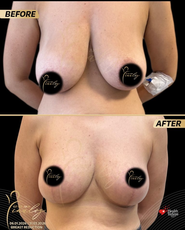
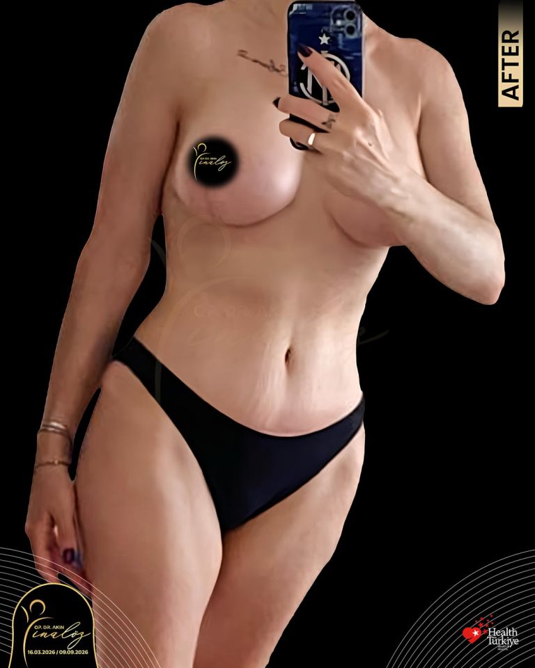
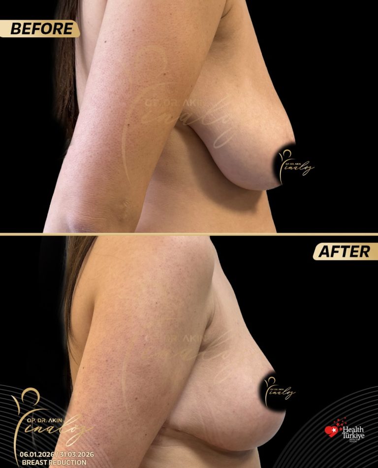
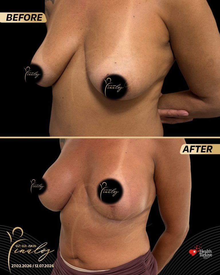
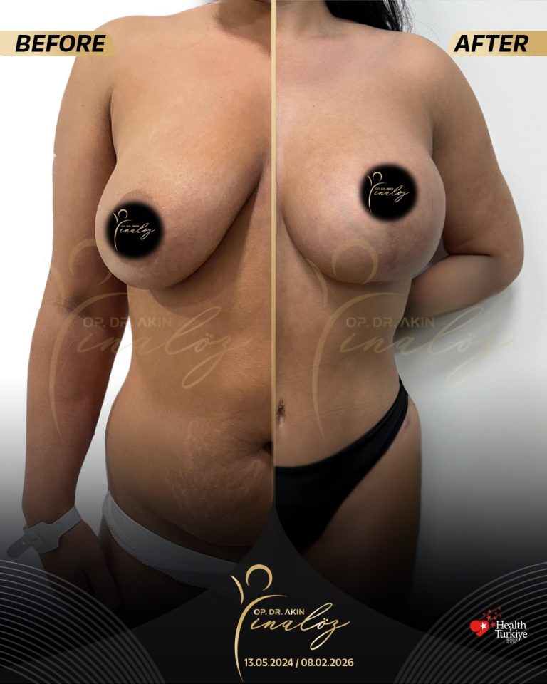
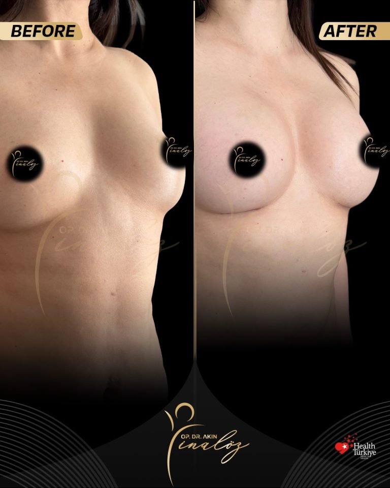
My goal is not just to perform a procedure, but to guide each patient through a safe, well-planned process toward a result that feels genuinely their own.
Results shown are of actual patients and may vary.
RELATED PROCEDURES
Considering Breast Surgery?
Contact info
- Optimist Residence, Fikirtepe, NO:10/35, 34720 Kadıköy/İstanbul
- +90 535 056 01 50
- info@akininaloz.com.tr