- Optimist Residence, Fikirtepe, NO:10/35, 34720 Kadıköy/İstanbul
- +90 535 056 01 50
- info@akininaloz.com.tr

BREAST LIFT
Comprehensive Guide to Breast Lift (Mastopexy) in Istanbul, Turkey
Breast lift surgery, clinically designated as mastopexy, is an advanced surgical intervention specifically engineered to elevate, reshape, and firm ptotic (sagging) breast tissue. Over time, physiological fluctuations, gravitational forces, systemic connective tissue laxity, structural hormonal shifts, and massive weight loss can compromise the elastic dermal envelope and internal suspensory ligamentous network, leading to the downward displacement of the mammary gland and the nipple-areolar complex (NAC). Performed within fully accredited surgical facilities in Istanbul, modern mastopexy protocols prioritize precise anatomical vector re-suspension, meticulous tissue redistribution, and predictable, aesthetically harmonious outcomes. International patients traveling from the United Kingdom and across Europe benefit from comprehensive surgical planning, high-grade surgical methodologies, and structured post-operative care pathways designed to ensure safety, clinical efficacy, and long-term aesthetic stability.
Modern aesthetic surgery has shifted definitively away from generalized standardization toward highly customized anatomical tailoring. Every thoracic structure exhibits unique biomechanical and morphological parameters, including chest wall width, skin envelope elasticity, native breast base diameter, soft-tissue thickness, and parenchymal distribution density. Achieving balanced, enduring results requires rigorous pre-operative planning, advanced surface mapping diagnostics, and an exhaustive understanding of soft-tissue mechanics under varying gravitational and muscular loads.
Anatomical Foundations and the Etiology of Ptosis
The structural integrity of the breast relies heavily on the superficial fascial system and the suspensory ligaments of Cooper, which anchor the glandular matrix to the deep pectoral fascia and the overlying dermal layer. When these supportive structures undergo elongation or degradation, the breast loses its natural upper-pole projection and youthful contour.
Regnault’s Classification of Ptosis: Pre-operative clinical evaluation categorizes the severity of sagging based on the relative position of the nipple-areolar complex compared to the inframammary fold. Grade I (mild) exhibits the nipple slightly below the fold; Grade II (moderate) places the nipple significantly lower but above the most dependent point of the breast tissue; Grade III (severe) repositions the nipple well below the lowest point of the breast mound, pointing directly toward the floor.
Parenchymal Redistribution and Auto-Augmentation: A primary objective of modern mastopexy is not merely skin excision, but the internal remodeling of the native glandular tissue. By shifting available parenchyma upward, surgeons can restore upper-pole fullness without necessarily introducing synthetic implants, though combined augmentation-mastopexy procedures remain an option for patients seeking both volume enhancement and elevation.
Surgical Modalities, Incision Patterns, and Technical Protocols
The execution of a mastopexy requires precise surgical planning regarding the exact incision pattern needed to excise redundant skin envelope structures while preserving the vascular and sensory integrity of the nipple-areolar complex.
1. Incision Patterns and Geometric Design
Periareolar (Donut) Mastopexy: Utilized primarily for mild ptosis or minor contour refinements, this circular incision is placed exclusively along the outer margin of the pigmented areola. It allows modest vertical lifting and diameter reduction but offers limited skin tightening capacity.
Vertical (Lollipop / Round Block) Mastopexy: Designed for moderate ptosis, this technique incorporates a periareolar incision combined with a single vertical line extending downward from the lower edge of the areola to the inframammary fold. It provides robust vertical elevation and superior tissue shaping capabilities.
Inverted-T (Anchor) Mastopexy: Indicated for severe ptosis, massive weight loss transformations, or large, heavy breasts requiring extensive tissue reduction and skin excision. The anchor pattern includes a periareolar line, a vertical component, and a horizontal closure hidden neatly within the inframammary crease, ensuring optimal long-term support.
2. Vascular Pedicle Preservation
Maintaining the delicate neurovascular supply to the nipple-areolar complex is a critical technical priority. Surgeons utilize specialized pedicle designs—such as superior, superomedial, or inferior dermal-glandular pedicles—to safely anchor and transpose the NAC to its newly elevated anatomical position while safeguarding sensory pathways and lactiferous duct viability.
The Comprehensive Clinical Pathway for International Patients
Undergoing advanced surgical procedures abroad requires a structured, multi-phase framework ensuring clinical continuity from initial virtual consultation to final post-operative clearance and long-term monitoring.
Remote Diagnostics and Initial Assessment: Initial evaluations involve digital photographic analysis, comprehensive medical history reviews, and virtual consultations to discuss anatomical expectations, review historical imaging, and establish preliminary surgical candidacy.
Pre-Operative Evaluation in Istanbul: Upon arrival at the accredited facility in Istanbul, physical examinations, comprehensive blood panels, and age-appropriate imaging (such as ultrasound or diagnostic mammography) are conducted by the multidisciplinary medical team.
Surgical Execution and Anesthesia Protocols: Interventions are performed under general anesthesia administered by qualified, board-certified anesthesiologists within sterile operating theaters equipped with advanced life-support and monitoring technology.
Immediate Post-Operative Monitoring: Patients are transferred to specialized recovery units for continuous vital sign tracking, pain management optimization, and early mobilization protocols under direct medical supervision.
Recovery and Follow-Up Protocols: Post-surgical check-ups verify proper wound healing, assess edema reduction, and clear the patient for safe return travel to the United Kingdom, supplemented by remote telemedical monitoring channels.

Op. Dr. Akın İnalöz
Plastic & aesthetic surgeon, Kadıköy, Istanbul
Quick Enquiry
Things You Might Be Wondering
Frequently Asked Questions
Most patients require approximately 7 to 10 days of local rest in Istanbul before undertaking international air travel or resuming sedentary work duties. Complete resolution of post-operative edema and the resumption of high-impact physical exercise or heavy lifting generally requires 4 to 6 weeks, subject to individual tissue healing characteristics.
Yes. An augmentation-mastopexy combines tissue elevation with volumetric enhancement. This combined approach restores youthful parenchymal positioning while simultaneously increasing upper-pole fullness, executed in a single surgical setting.
While advanced pedicle preservation techniques aim to protect the lactiferous ducts and nerve pathways, any major breast surgery carries a variable risk of impacting milk production. Patients planning future pregnancies should discuss this thoroughly during their comprehensive pre-operative consultation.
Surgeons utilize meticulous tension-free multilayer closure techniques, dissolvable subcutaneous sutures, and specialized atraumatic instrumentation. Post-operative scar management protocols, including silicone sheeting and topical silicone gels, are subsequently recommended during the maturation phase.
While mastopexy establishes a long-lasting, youthful contour baseline, natural aging, gravitational forces, and significant weight fluctuations can gradually influence tissue elasticity over subsequent years. Maintaining a stable lifestyle helps preserve the surgical outcome indefinitely.
Travelling from the UK for breast surgery?
See how the consultation, treatment and recovery process works, start to finish.
Op. Dr. Akın İnalöz
Natural Aesthetic Evolutions
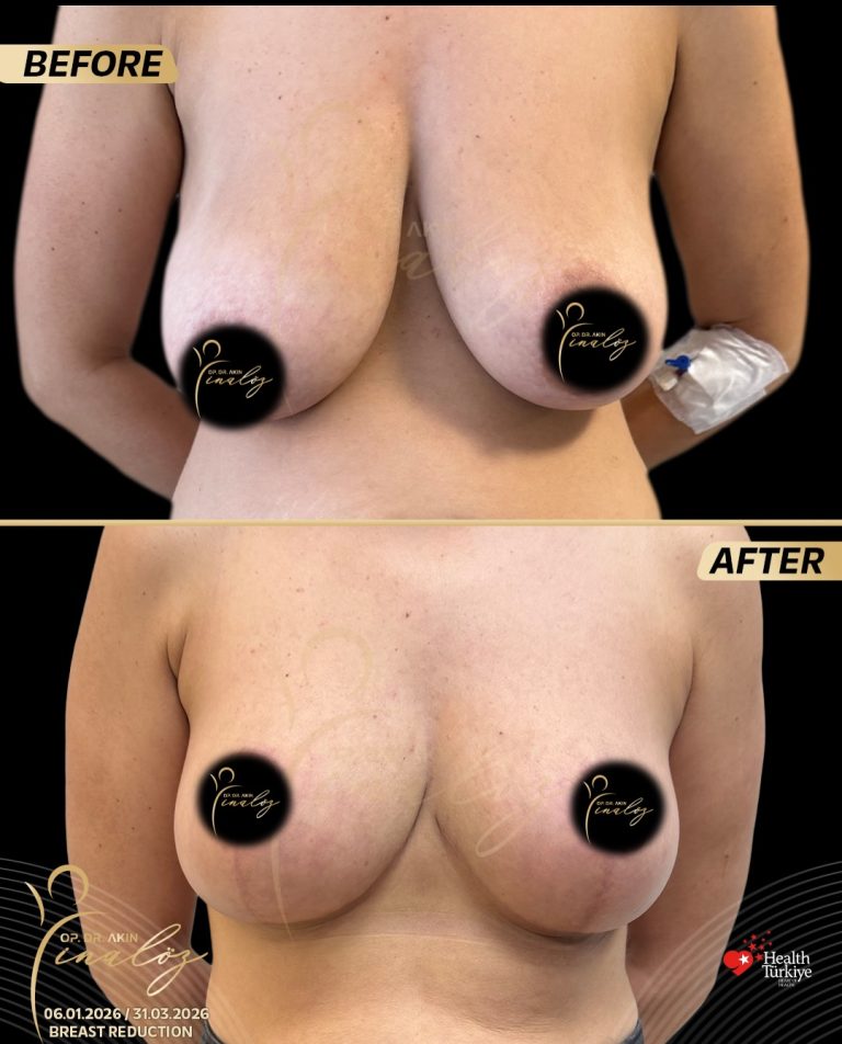
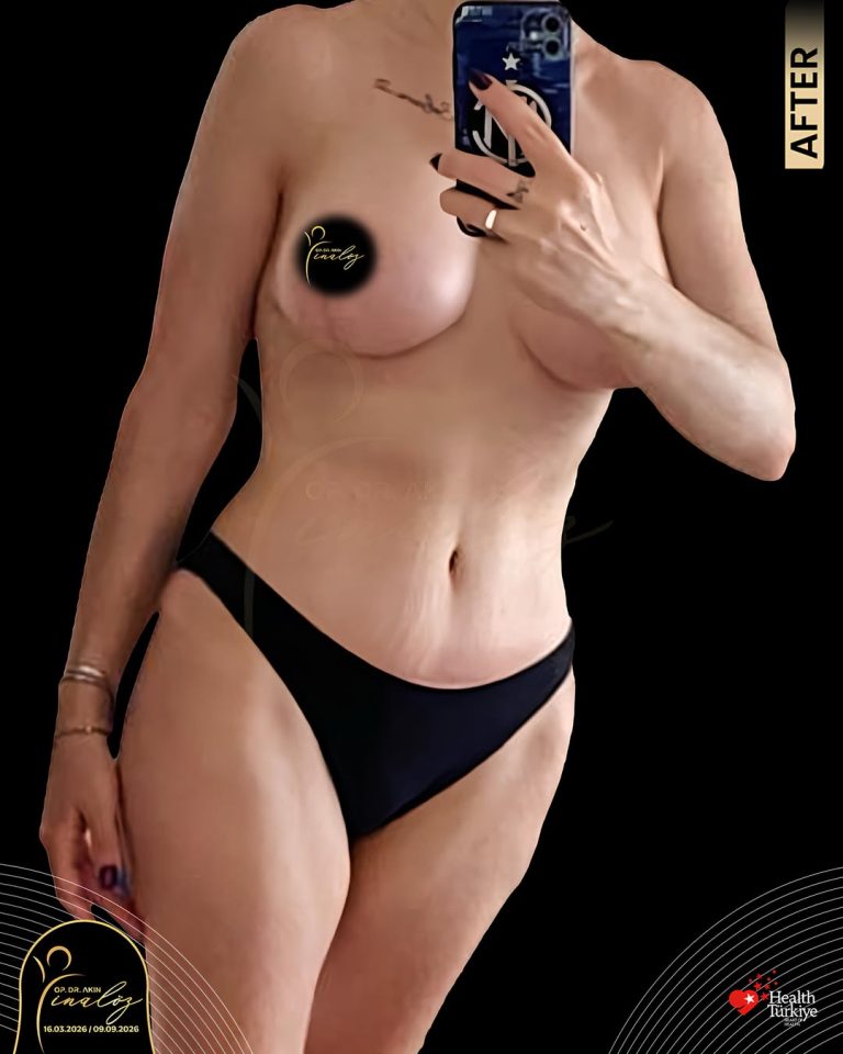
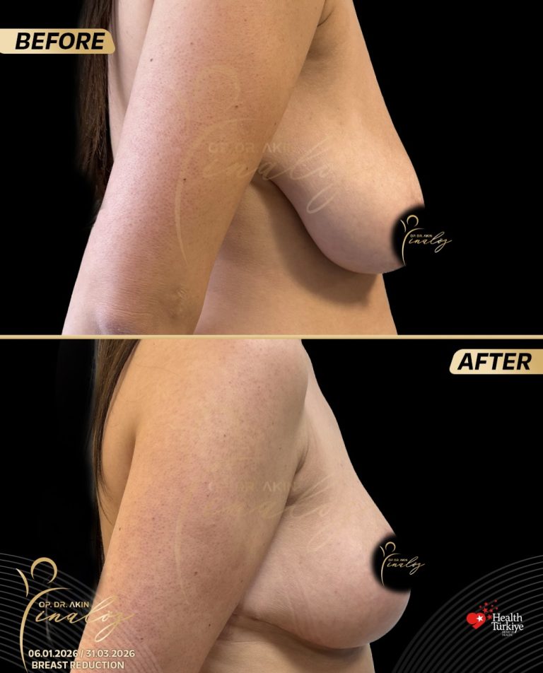
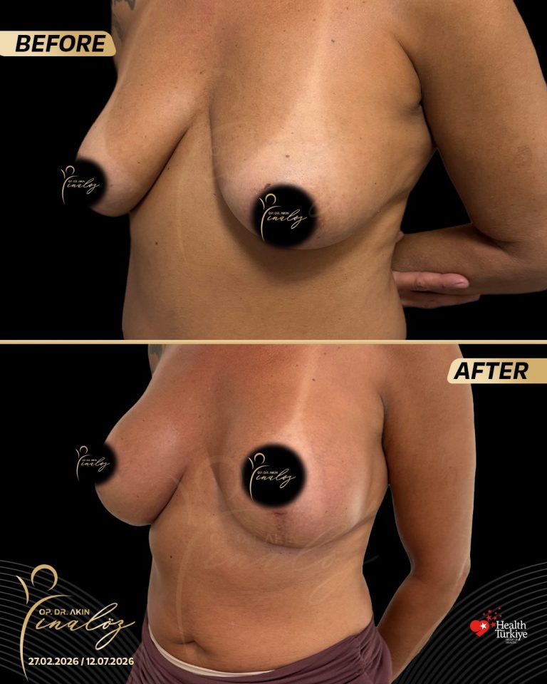
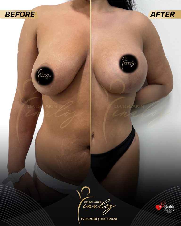
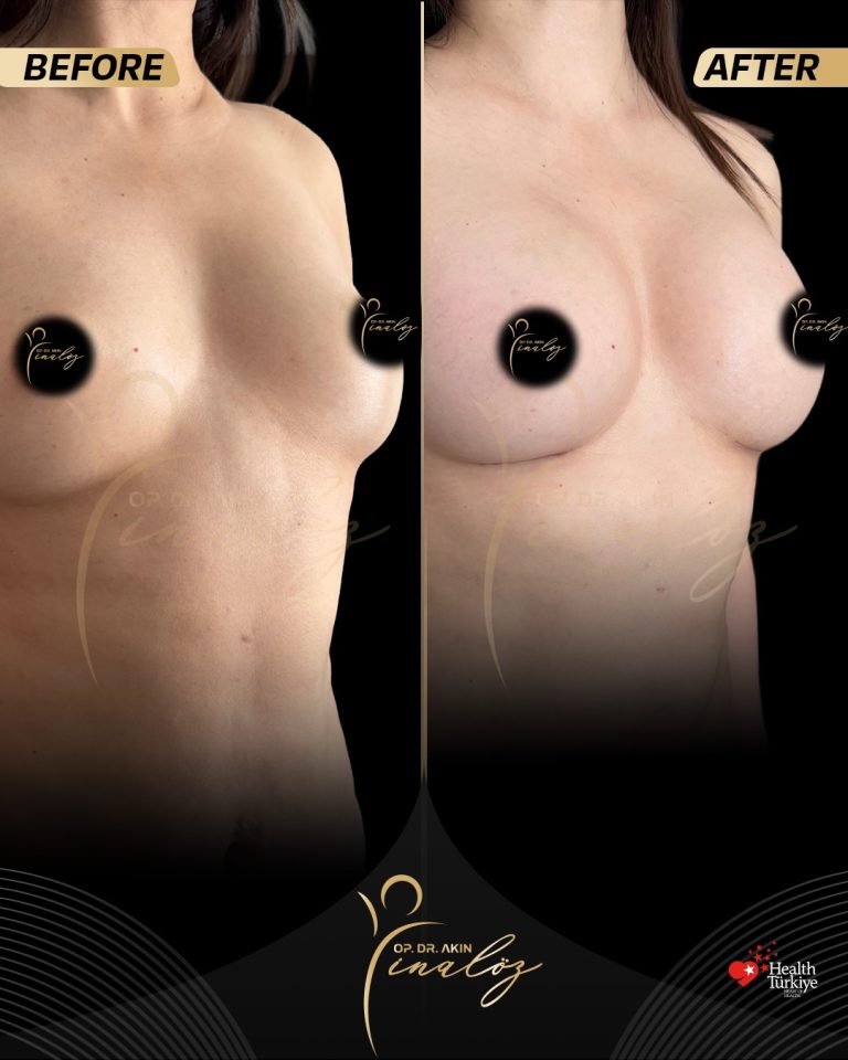
My goal is not just to perform a procedure, but to guide each patient through a safe, well-planned process toward a result that feels genuinely their own.
Results shown are of actual patients and may vary.
RELATED PROCEDURES
Considering Breast Surgery?
Contact info
- Optimist Residence, Fikirtepe, NO:10/35, 34720 Kadıköy/İstanbul
- +90 535 056 01 50
- info@akininaloz.com.tr